4 Chambers
- Left atrium (7)
- Left ventricle (8)
- Right atrium (2)
- Right ventricle (3)
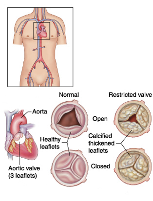
4 Valves
- Aortic valve (1)
- Mitral valve (2)
- Tricuspid valve (3)
- Pulmonary valve (4)
The heart is a muscle that pumps oxygen rich blood (red blood) and nutrients to all parts of your body through tubes called arteries. Like all muscles in the body, the heart also needs oxygen to work efficiently. Oxygen is supplied by the coronary arteries. When these arteries become significantly narrowed, less oxygen is supplied and chest pain on exertion can develop. This is called angina. If the coronary arteries block completely then a heart attack occurs and the muscle area supplied by this artery then dies. The blocked or narrowed arteries can be reopened using a balloon and a metal tube called a stent. This procedure is called angioplasty (see case 1).
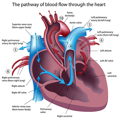
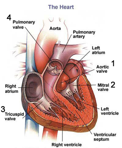
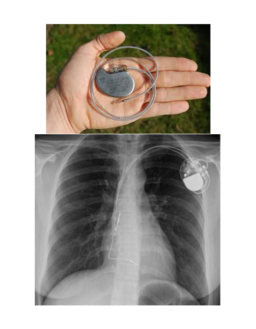
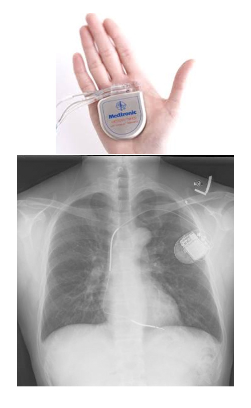
The heart has four chambers (the right atrium, the left atrium, the right ventricle and the left ventricle – figure 1). Each heart beat is a result of an electrical stimulation that starts in the top right hand chamber of the heart (the right atrium) and this electrical impulse is conducted down “cables” to the various chambers (image). Any part of this electrical circuit can become diseased resulting in either slow or fast heart rates. Either very slow or very fast heart rates can result in palpitations, dizziness or a collapse (loss of consciousness). If the heart is too slow then a pacemaker is required (figure 2). If the heart rate is too fast, then depending on the mechanism of the fast heart rate, either tablets, ablation therapy (using heat energy to modify some of the electrical circuits) or a defibrillator (like a pacemaker but treats very fast life threatening heart rhythms), may be required (figure 3).
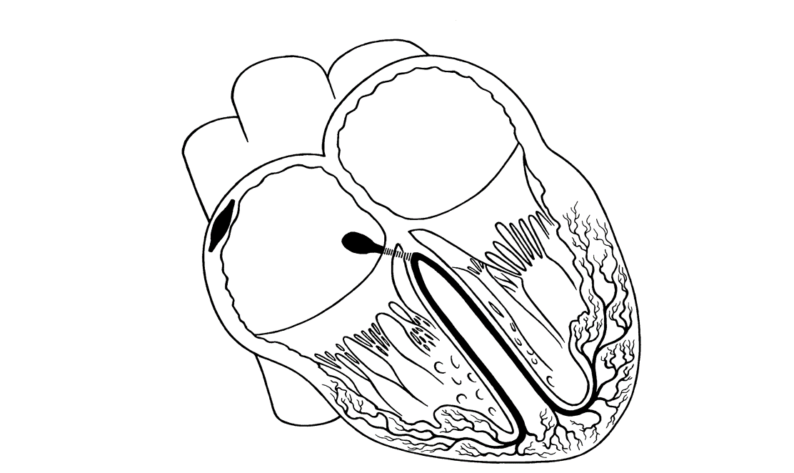
The heart has four valves (figure 1). Two on the left side of the heart (the mitral valve and the aortic valve), and two on the right side (the tricuspid valve and the pulmonary valve). Any of these valves can be affected but the commonest valves to become diseased are the mitral valve or the aortic valve. These valves can either be leaky (regurgitation) or become narrowed (stenosis). Patients can present with symptoms of shortness of breath, chest pain/discomfort or palpitations. A diseased valve (figure 4) creates turbulent flow as blood passes over it. This turbulent flow result in a sound called a murmur, which can be heard with a stethoscope.
When the pumping action of the heart fails, a condition called heart failure develops. This can result in symptoms of shortness of breath, fatigue and swollen ankles. The treatment of this condition is dependent of the cause of the injury to the heart muscle.
The heart, like all muscles in the body can enlarge or thicken when overworked. When the blood pressure is too high (hypertension or high blood pressure) this can occur. Hypertension can result is strokes, heart failure, kidney failure and heart attacks. Some patients can also develop a thicken heart muscle even though their blood pressure is normal. This condition is called hypertrophic cardiomyopathy. Patients with this condition may have a family history of sudden unexpected death.